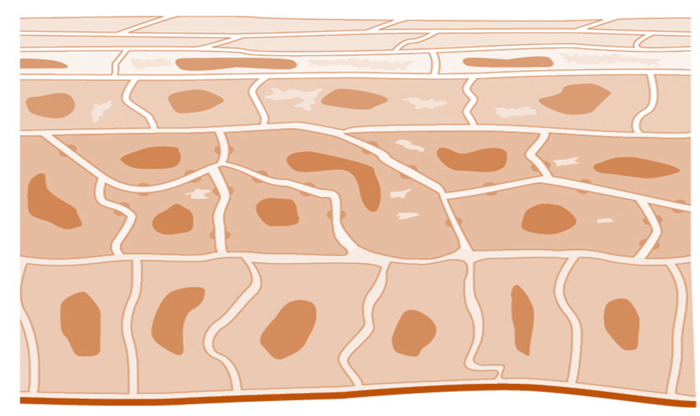
Keratinocyte Structure and Function

Keratinocyte cells are found in the deepest basal layer of the stratified epithelium that comprises the epidermis, and are sometimes referred to as basal cells or basal keratinocytes. It is known that 95% of the cells in the epidermis are keratinocytes. Squamous keratinocytes are also found in the mucosa of the mouth and esophagus, as well as the corneal, conjunctival and genital epithelia.
Keratinocytes are maintained at various stages of differentiation in the epidermis and are responsible for forming tight junctions with the nerves of the skin. They also keep Langerhans cells of the epidermis and lymphocytes of the dermis in place.
Immune Role of Keratinocytes
In addition to their structural role, keratinocytes play a role in immune system function. The skin is the first line of defense and keratinocytes serve as a barrier between an organism and its environment. In addition to preventing toxins and pathogens from entering an organisms body, they prevent the loss of moisture, heat and other important constituents of the body. In addition to their physical role, keratinocytes serve a chemical immune role as immunomodulators, responsible for secreting inhibitory cytokines in the absence of injury and stimulating inflammation and activating Langerhans cells in response to injury. Langerhans cells serve as antigen-presenting cells when there is a skin infection and are the first cells to process microbial antigens entering the body from a skin breach.
Differentiation of Keratinocytes
Keratinocyte stem cells reside in the basal layer of the epidermis, which is the lowest layer of the stratified epithelia. These cells divide to give rise to transient amplifying cells which divide further, and differentiate, as they move upwards in the epidermis. The differentiating cells produce compounds and other proteins which are critical to the integrity of the outermost layer of the skin, the stratum corneum. The keratinocytes in the stratum corneum are dead squamous cells that are no longer multiplying. Once keratinocytes reach the corneum, they are said to be keratinazed, or cornified, creating the tough outer layer of skin.
The major proteins found in keratinocytes are keratins. These proteins form the cytoskeleton of keratinocytes, and keratin expression changes as transient amplifying cells differentiate and move to the most superficial stratum corneum. These keratins are what make up our hair and nails, which is why defects in keratin expression result in various diseases of the epidermis, as well as the hair and nails.
Links
CRO Pre-clinical Research Services: Xenograft animal models
Generation of Stably Expressing Cell Lines in 28 Days
Stable RNAi Cell Line Generation: Stable Gene Knockdown
In Vivo RNAi: Tissue-targeted siRNA
Encapsulation of Protein, RNA, mRNA, and DNA Molecules into Liposomes
siRNA Delivery – In Vivo Transfection Kits
General Information | Keratinocytes of the Skin | Cell Culture | Keratinocyte Transfection Kit